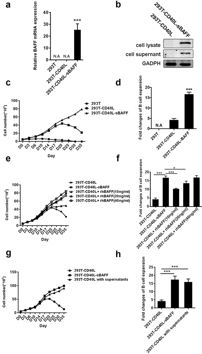
Figure 1. The 293T-CD40L-sBAFF cell line had the better potential to stimulate B cell growth.

a. BAFF mRNA expression was confirmed by quantitative RT-PCR. b. Western-blotting confirmed the presence of Flag-tagged soluble BAFF in the cell lysate and the supernatant. c-h. B cells from multiple healthy adult donors were co-cultured with the irradiated 293T, 293T-CD40L or 293T-CD40L-sBAFF feeder cells in the presence of cytokine cocktails as well as the indicated treatments. The B cells were harvested every 3-5 days and the cell numbers excluding trypan blue-positive dead cells were counted, with the average measurements for three donors and their standard deviations over time are shown. (c) Expansion patterns and (d) The B-cell expansion change in the presence of 293T-CD40L-sBAFF cells, 293T-CD40L cells, or 293T cells at day 35. (e) Expansion patterns and (f) expansion fold change of B-cells in the presence of 293T-CD40L-sBAFF cells, or 293T-CD40L cells with varying concentrations of recombinant human BAFF (rhBAFF) at day 35. (g) Expansion patterns and (h) expansion fold change of CD40L-B cells with the filtered supernatants of 293T-CD40L-sBAFF cells at day 35. Data represent mean ± SD (error bars) for a representative experiment of n = 3 independent experiments. The paired t-test and one-way ANOVA were used. P < 0.05 indicates statistically significance difference. * indicates P < 0.05; ** indicates P < 0.01; *** indicates P < 0.001.
