
Figure 5. EPHB6 action is mediated by the ERK2 kinase.
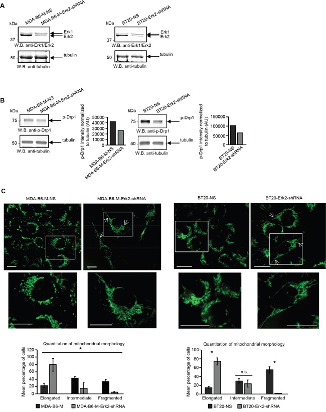
A. MDA-B6-M and BT-20 cells were transiently transduced with ERK2-targeting shRNA (MDA-B6-M-ERK2-shRNA and BT20-ERK2-shRNA) or non-silencing shRNA (MDA-B6-M-NS and BT20-NS) and ERK2 expression was examined by Western blotting. B. Phosphorylation of DRP1 was analysed in the indicated cells by Western blotting. DRP1 phosphorylation was quantitated by densitometry, measurements were normalized to matching tubulin controls and are presented in arbitrary units (AU). C. Mitochondria were visualised in the indicated cells by confocal microscopy as in Figure 2A. Arrows indicate elongated mitochondria. Selected areas are shown at higher magnifications. Z-stack frames were split and fluorescence intensity was adjusted using the Fiji software. Scale bar, 20 μm. Quantitative analysis was done as in Figure 2A. Each graph represents two independent experiments. * p<0.05, Student's t-test. n.s., statistically not significant. All experiments were repeated at least twice.