
Figure 5. Flow cytometric analysis of cell cycle progression in human pancreatic cancer cells treated with PA-MSHA.
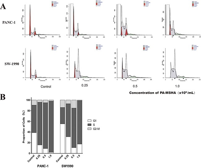
A. The columns represent the mean ± SD of the three independent experiments. PANC-1 and SW-1990 were treated with the indicated concentrations of PA-MSHA in serum-free medium for 24 h. The results are representative of three independent experiments. B. The percentages of cells in G1, S, and G2-M are shown as histograms