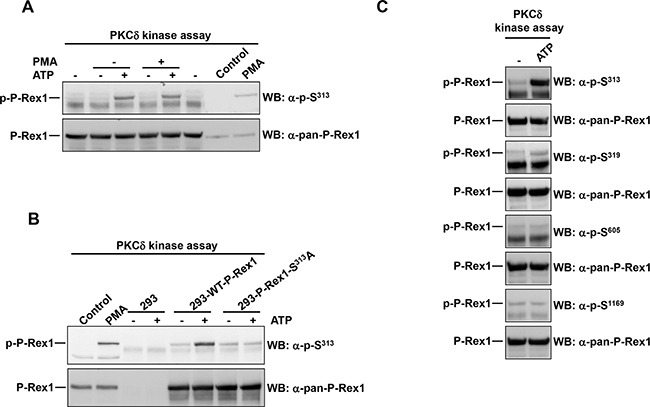
Figure 4. PKCδ phosphorylates P-Rex1 at Ser313.

A. P-Rex1 immunoprecipitated from extracts of MCF7 cells was used as substrate in a PKCδ in vitro kinase assay. ATP or PMA were added in the kinase assay where indicated. After completion of the kinase assay, P-Rex1 pS313 and total P-Rex1 levels were analyzed by Western blot. B. P-Rex1 was immunoprecipitated from extracts of 293 cells transfected with wild type P-Rex1 or P-Rex1 mutated in serine 313 to alanine and used as substrate in PKCδ in the in vitro kinase assay. The levels of P-Rex1 pS313 and total P-Rex1 were analyzed by Western blot. C. MCF7 cells were lysed and the extracts were used to immunoprecipitate P-Rex1. Then the PKCδ kinase assay was performed. P-Rex1 phosphorylation levels in serine 313, 319, 605, 1169 and total P-Rex1 were analyzed by Western blot with anti-phospho-specific and anti-pan-P-Rex1 antibodies. The blots shown come from an experiment that was repeated at least twice.
