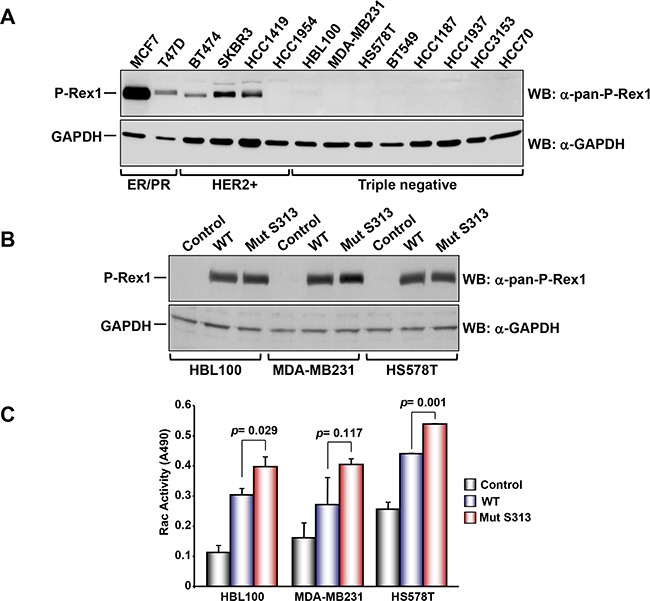
Figure 5.

A. Analysis of the expression of P-Rex1 in different breast cancer cell lines. The different cells lines were lysed and the expression of P-Rex1 was analyzed by Western blot. B. HBL100, MDA-MD231 and HS578T cells were transfected with vectors coding for wild type P-Rex1 or P-Rex1 mutated in S313A and lysed. The expression of the different forms was analyzed by Western blot with the anti-pan-P-Rex1 antibody. GAPDH was used as loading control. C. Rac activity in the cell lines described above and transfected with wild type P-Rex1 or P-Rex1 mutated in S313A. The blots shown come from an experiment that was repeated at least twice.
