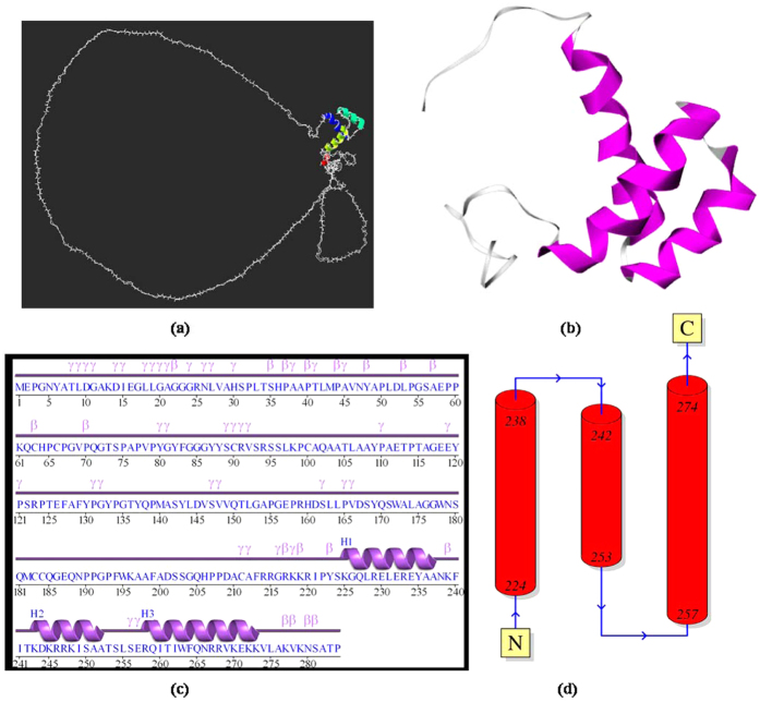
Figure 6. Validation results of the hoxb13.B99990026.pdb modelled protein by PDBsum.
(a) 3D protein structure of the complete HOXB13 protein (hoxb13.B99990026.pdb) modelled using MODELLER v9.17 (Figure generated using SWISSPDBViewer). (b) HOXB13 (2CRA) protein 3D structure (Figure generated using PDBsum). (c) The amino acid residues contributing to the secondary structure (alpha helix and beta turns) of the complete HOXB13 protein are depicted in the topology diagram (Figure simulated using PDBsum). (d) Linear view of the modelled complete HOXB13 protein structure with alpha helices, beta and gamma turn and corresponding amino acid residues (Figure simulated using PDBsum).

