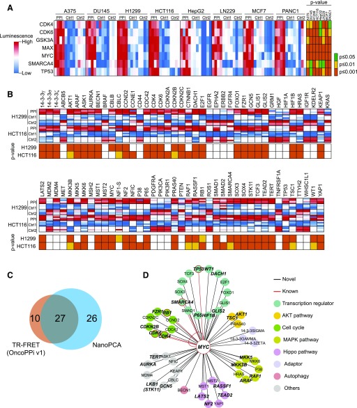
Fig. 4.
High-throughput PPI mapping of oncogenic MYC interactome. (A) Heat map of known MYC PPIs in eight cell lines showing four independent replicates. (B) Oncogenic PPI profiling of N-NLuc-MYC against C-NLuc–tagged genes library. Four independent replicates were shown in the heat map. (C) The Venn diagram of the distribution of positive PPIs identified in NanoPCA platform and TR-FRET–based OncoPPi v1 network (Li et al., 2016). (D) An oncogenic MYC PPI hub. PPIs that are positive with S/B > 1.0 and P < 0.05 in both cell lines were selected. Colors are assigned as indicated. PPIs that are double positive in both NanoPCA and TR-FRET were denoted in bold italic. MAPK, mitogen-activated protein kinase.

