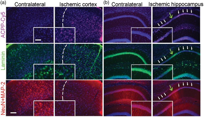
Figure 5.
Uptake of ACPP-Cy5 is associated with loss of neuronal laminin and neurodegeneration in ischemic brain. ACPP-Cy5 (2 nmol) was injected i.v. after 2-h filament-induced MCA occlusion in mice, which were sacrificed 24 h later. Sections were immunostained with the neuronal markers NeuN and MAP-2 (red) to visualize cell bodies and processes; ECM laminin was labeled with anti-laminin antibody (green) and cell nuclei with Hoechst dye (blue). (a) Neuronal loss (bottom panel) and adjacent laminin staining in the vasculature (middle panel) correlate with uptake of ACPP-Cy5 (cyan, top panel) in the ischemic cortex (separated by a white dashed line). (b) In ischemic hippocampus, white arrows indicate areas of increased ACPP-Cy5 uptake (top panel), correlating with loss of laminin immunoreactivity (middle panel) and neuronal degeneration (loss of neuronal markers in bottom panel). In contrast, green arrows indicate areas of decreased ACPP-Cy5 uptake, correlating with relatively intact laminin and less impaired neuronal processes. The contralateral hippocampus manifested minimal ACPP-Cy5 uptake. Scale bar, 30 µm (top, inset) and 75 µm (bottom).

