Fig. 9.
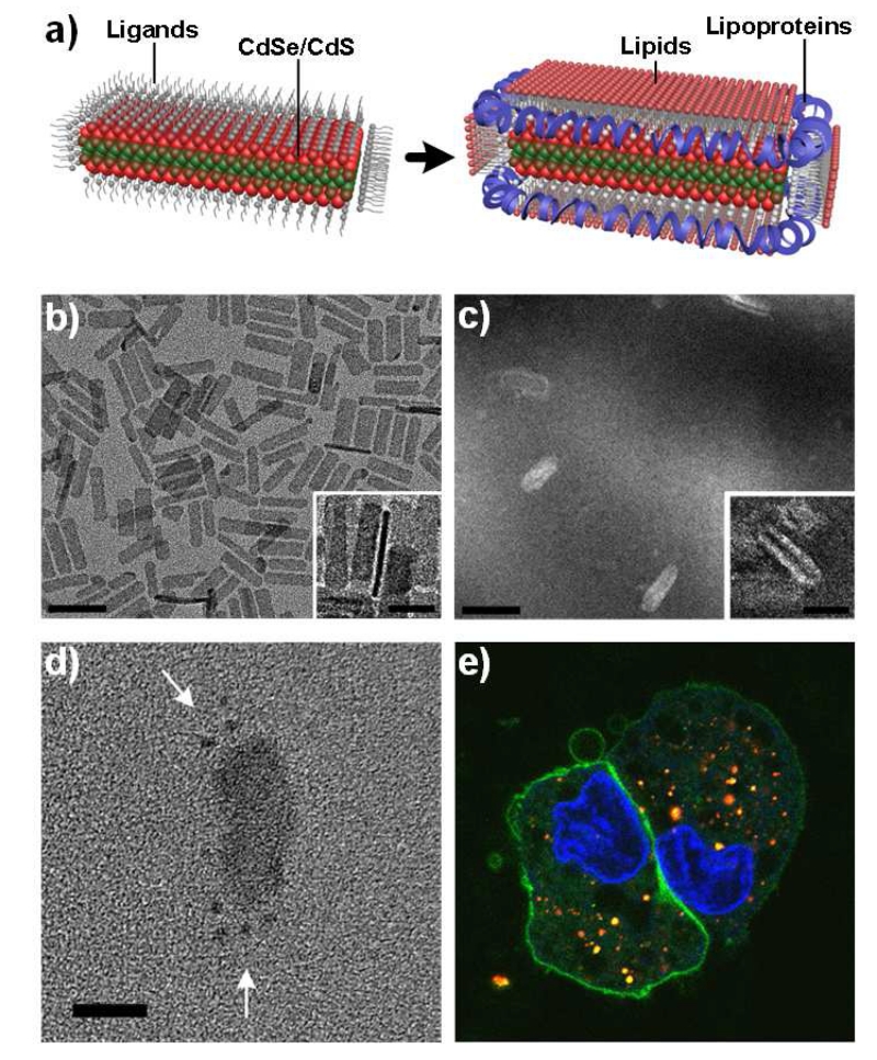
Micellar encapsulation of sheet-like nanoplatelets using a combination of lipids and membrane scaffold proteins (MSP). Both coatings are required to enable full coating of flat surfaces and highly curved surfaces of the nanoplatelets. (a) Schematic depiction of the nanoparticle coating process. (b) Transmission electron microscopy image of CdSe/CdS core/shell nanoplatelets. (c) Counterstained electron microscopy image of encapsulated nanoplatelets. Insets show side-views of particles. Scale bars = 50 nm in wide-field images and 20 nm in insets. (d) Encapsulated nanoplatelets after MSP conjugation to spherical gold colloids to localize the protein with respect to the nanoplatelet. Arrows indicate the gold. Scale bar = 50 nm. (e) Fluorescence image of A431 cells 24 hours after exposure to lipoprotein-nanoplatelets, shown in red. Cells are counterstained with a nuclear dye (blue) and a plasma membrane dye (green). Nanoplatelets remain brightly fluorescent and localize throughout the cells. Image reproduced with permission from reference [195].

