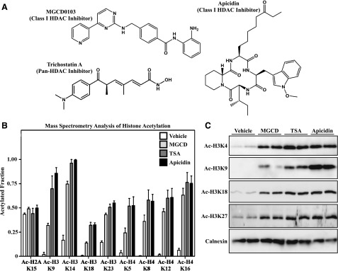
Fig. 1.
Effects of structurally distinct HDAC inhibitors on histone acetylation in cardiac fibroblasts. (A) Chemical structures of MGCD0103, TSA, and apicidin. (B) Site-specific changes in acetylation of lysine residues in histones H2A, H3, and H4 were quantified by mass spectrometry using lysates from AMVFs treated with DMSO vehicle control (0.1% final concentration), MGCD0103 (1 μM), TSA (1μM), or apicidin (3 μM) for 24 hours. Data are expressed as the fraction of acetylated versus nonacetylated residues ±S.E.M. (n = 4/group). (C) AMVFs were treated with HDAC inhibitors for 24 hours, homogenized, and subjected to immunoblotting with the indicated anti–acetyl-histone antibodies. Calnexin served as a loading control. Each lane represents an independent plate of cells. MGCD, MGCD0103.

