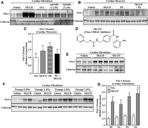
Fig. 3.
Benzamide HDAC inhibitors stimulate PAI-1 protein expression in cardiac fibroblasts. (A) Immunoblot analysis of PAI-1 expression in cultured, passage 2 ARVFs treated with DMSO vehicle, MGCD0103 (1 μM), TSA (200 nM), or apicidin (1–3 μM) for 24 hours. (B) Treatment of neonatal rat ventricular cardiomyocytes for 48 hours with MGCD0103 in the absence or presence of the hypertrophic agonist PE (10 μM) failed to enhance PAI-1 expression. (C) The PAI-1 signal in (B) was quantified by densitometry and normalized to the loading control, calnexin. Data represent means ± S.E.M. (differences were not statistically significantly different). (D) Chemical structure of the benzamide class I HDAC inhibitor, MS-275. (E) Treatment of ARVFs with MGCD0103 (1 μM) or MS-275 (1 μM) for 24 hours led to increased PAI-1 protein expression. (F) Passage 0, 1, 2, or 3 ARVFs were treated with MGCD0103 (1 μM) for 16 hours. The ability of MGCD0103 to increase PAI-1 expression was unaffected by myofibroblast phenotype, as defined by the progressive increase in α-smooth muscle actin expression upon fibroblast passage. For all blots, each lane represents an independent plate of cells. (G) The PAI-1 signal in (F), as well as from other samples from an independent preparation of ARVFs (not shown), was quantified by densitometry and normalized to the loading control, calnexin (n = 5 plates of cells/condition). Data represent means ± S.E.M. (*P < 0.05 versus vehicle-treated cells). MGCD, MGCD0103; PE, phenylephrine; Veh, vehicle.

