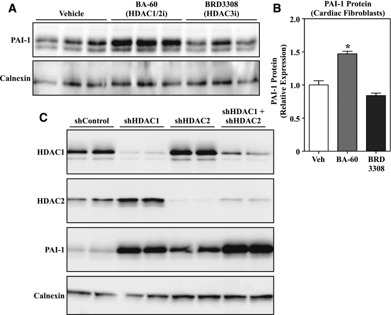
Fig. 5.
MGCD0103-induced PAI-1 expression is mediated by HDAC1 and HDAC2 inhibition. (A) Western blot analysis of PAI-1 expression in cultured ARVFs treated with the isoform-selective HDAC inhibitors BA-60 (HDAC1/2 inhibitor, 300 nM) and BRD3308 (HDAC3 inhibitor, 1 μM) for 24 hours. (B) The PAI-1 signal in (A) was quantified by densitometry and normalized to the loading control, calnexin. Data represent means ± S.E.M. (*P < 0.05 versus vehicle-treated cells). (C) AMVFs were infected with shControl or lentiviruses encoding shRNAs to target HDAC1 and/or HDAC2. After 96 hours of infection, cell homogenates were subjected to immunoblotting with antibodies specific for HDAC1, HDAC2, and PAI-1. Calnexin served as a loading control. Each lane represents an independent plate of cells. BA-60, biaryl-60; shControl, short-hairpin control lentivirus; shRNA, short-hairpin RNA; Veh, vehicle.

