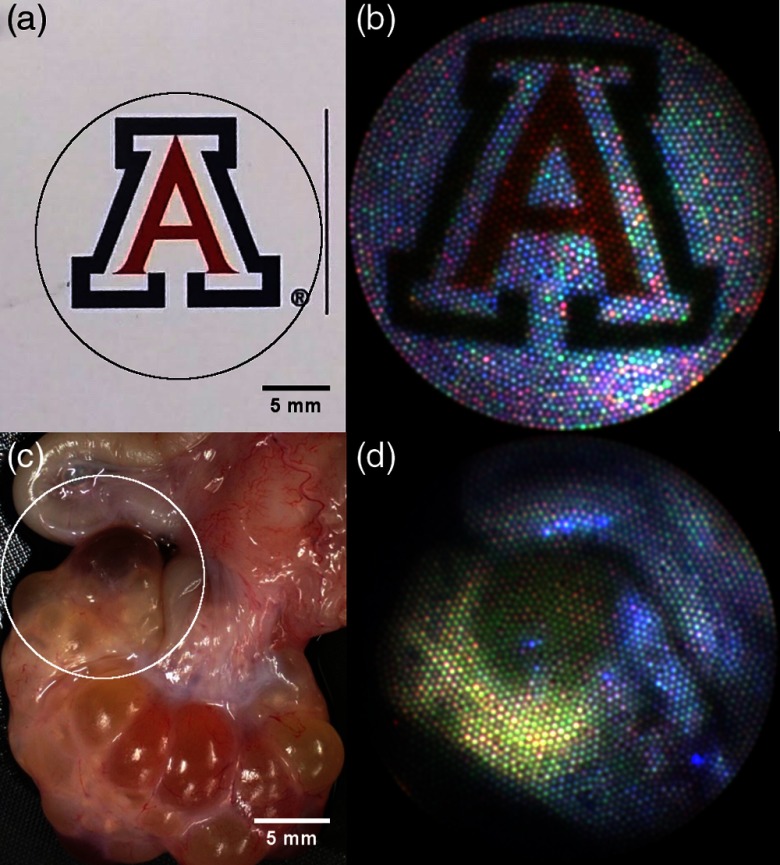
Fig. 7.
Images on left are taken using a digital camera and are of (a) the University of Arizona logo on a business card and (c) a section of a porcine reproductive system including ovary with follicles (bottom) and fallopian tube (top left). Corresponding images taken with the endoscope are on the right with FOV approximated by circles overlaid on left images. Image B is a composite color reflectance images taken by sequentially acquiring images with 405-, 515-, and 642-nm illumination. Image D is a composite color fluorescence image created using 375-nm excitation and sequentially acquiring images using the 450-, 532,- and 590-nm emission filters.

