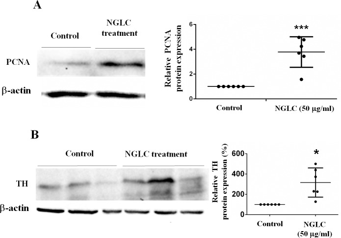
Fig 5. Proliferating cell nuclear antigen (PCNA) and thyroxin hydroxylase (TH) western-blots analysis.
SN4741 cells were cultured for 7 days with a high concentration (50 μg/ml) of nanocrystalline glass-like carbon (NGLC) microflakes. PCNA (A) and TH (B) analysis for relative protein expressions were expressed as percentage. The data are shown as mean± standard error of the means (SEM, n = 3) and Student’s t tests were used for statistical significance between two groups; *p<0.05 and ***p<0.001.

