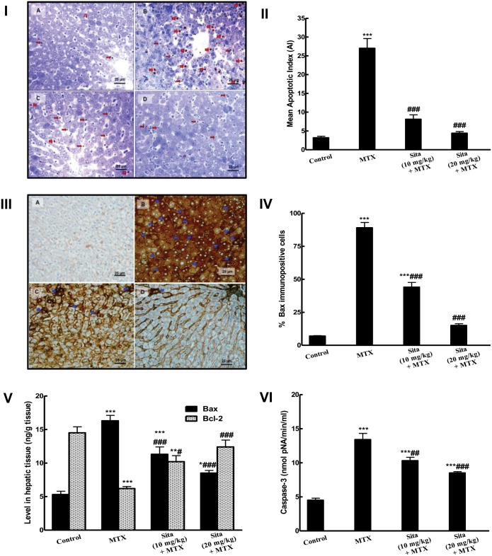
Fig 5. Sitagliptin (Sita) pretreatment counteracts apoptosis and improves apoptotic markers in the hepatic tissues of Methotrexate (MTX)-treated mice.
I. TUNEL-positive apoptotic liver cells nuclei (Thick red arrow) and bodies (Thin red arrow) detected in the liver sections were: (A) Occasionally seen in Control mice group; (B) Frequently observed in mice group intoxicated with MTX; (C&D) Clearly decreased in sita pretreated groups with more marked reduction in the number of apoptotic cell and bodies seen in group pretreated with sita 20 mg/kg. II. Effect on mean apoptotic index (the number of TUNEL-positive cells/high- power field (× 400) across 10 different fields for each TmA section). III. The expression of pro-apoptotic marker Bax by immunohistochemical staining (400×): A) Control group with focal light brown cytoplasmic immunostaining in less than 1% of cells; B) MTX group, showing intense brown cytoplasmic immunostaining of Bax (Blue arrows) in more than 95% of cells (C, D) Sita + MTX groups, showing a marked decrease in the cytoplasmic expression of Bax in a dose dependent manner of sita. IV. Semiquantitative analysis of Bax immunohistochemical staining results in liver tissues of different groups, expressed as % of immunopositive cells in TmA sections of all animals of each group, 10 different fields/section. V. Levels of Bax and Bcl2 in hepatic tissue. VI. Caspase activity in hepatic tissue. n = 10. *P < 0.05, **P < 0.01, ***P < 0.001 vs. the control. #P < 0.05, ##P < 0.01, ###P < 0.001 vs. the MTX group (ANOVA followed by Tukey-Kramer multiple comparison).

