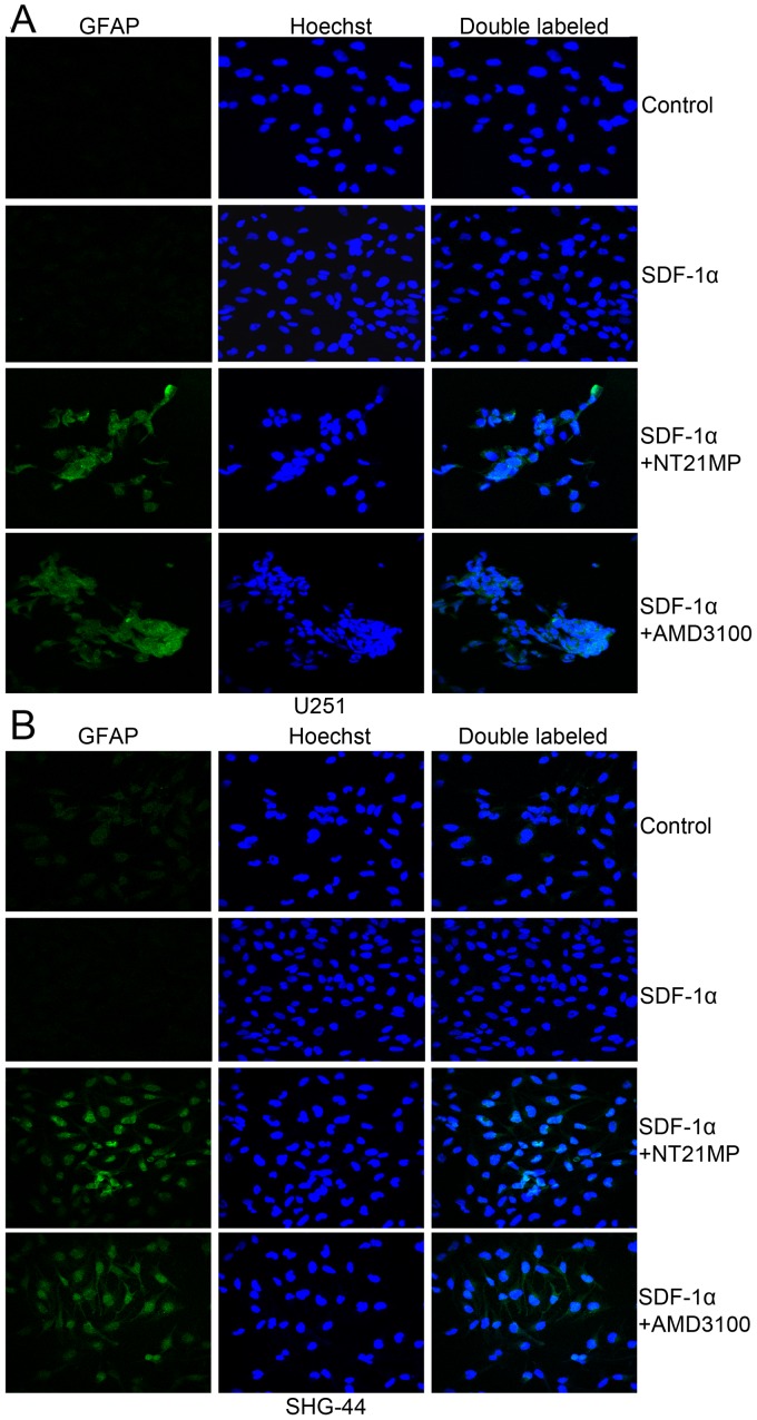
Figure 6.
Immunofluorescent staining of GFAP protein in U251 and SHG-44 cells. The cells were treated with (+SDF-1α) or not (−SDF-1α) with 100 ng/ml of SDF-1α and NT21MP (1 µg/ml) or AMD3100 (1 µg/ml). (A) The expression of GFAP in U251 cells. (B) The expression of GFAP in SHG-44 cells. Both images are at a magnification of ×200.

