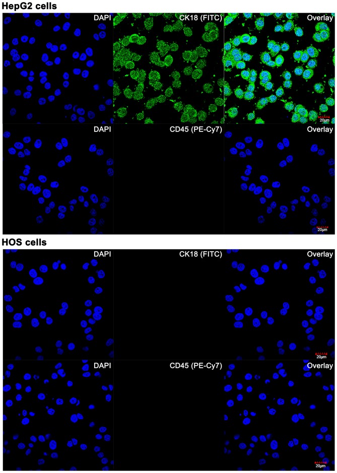
Figure 2.
Confocal laser scanning images of cultured HepG2 and HOS cells stained with fluorophore-conjugated antibodies against cytokeratin 18 (CK18, top panels) and CD45 (bottom panels). Nuclei were stained with DAPI. A Leica SP5 confocal system was used. DAPI was excited at λex = 405 nm (15% of maximum output power), FITC and PE-Cy7 were excited at λex = 488 nm (60% of maximum output power). For DAPI, emission was measured at λem = 461±50 nm (PMT at 467 V). For FITC and PE-Cy7, emission was measured at λem = 519±50 nm (PMT at 662 V). All parameters were kept constant for the imaging.

