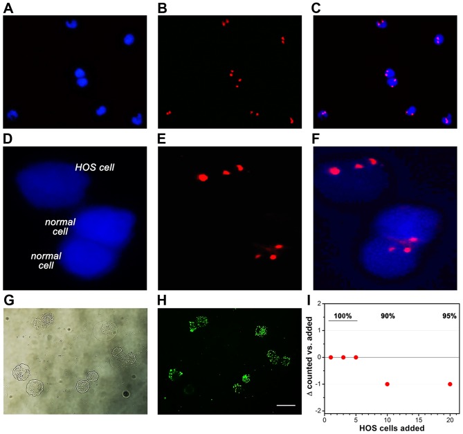
Figure 3.
Retrieval of epithelial and HOS cells derived from spiked peripheral blood samples of healthy volunteers. The top row shows cells that were retrieved from non-spiked blood (representative images of N=3 samples). Nuclei were stained with DAPI (blue fluorescence, A) and chromatin was stained with CEP 8 FISH (red fluorescence, B). The overlay is presented in (C). HOS cells in spiked peripheral blood could be distinguished from non-cancerous (CK18+/CD45−) cells (normal cell) on the basis of the aneuploidic chromatin number (D–F) (representative images of N=3 spiked samples). To determine retrieval efficiency of HOS cells using the isolation procedure employed for FISH, HOS cells were stained with the CYTO-ID green long-term cell tracer (brightfield in G and fluorescence in H) and added to 7.5 ml of peripheral blood to a final concentration of 1, 3, 5, 10, and 20 HOS cells/ml. Following isolation, the cells were counted by fluorescence microscopy in a 200-µl sample volume. (I) A modified Bland-Altman plot shows the retrieval efficacy of fluorescent HOS cells from the spiked PB samples. Retrieval rates are indicated as percentage at the top.

