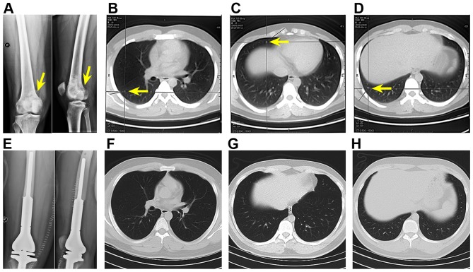
Figure 8.
X-ray and chest CT scans of a primary metastatic osteosarcoma patient. Upon OS diagnosis (A, arrow), there were three metastases in the right lung (B–D, arrows). After neoadjuvant chemotherapy, tumor segment resection, and prosthetic replacement (E), no metastases were observed on the chest CT scans (F–H, which correspond to the x-y cross-sectional planes where metastases were found before neoadjuvant chemotherapy and limb salvage surgery in B–D). Nevertheless, 4 CTCs were found in this patient's liquid biopsy.

