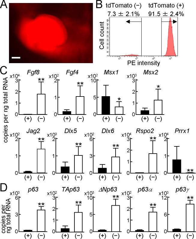
Fig 2. Expression of p63 variants in the AER and limb mesenchyme.
(A) Fluorescence image of a forelimb bud in a Prrx1-Cre;Rosa-CAG-LSL-tdTomato (Ai14) E11.5 embryo. Scale bar, 200 μm. (B) Flow cytometric analyses of forelimb bud cells from Prrx1-Cre;Ai14 E11.5 embryos. Error bars indicate s.d. (n = 3 biological replicates). (C) mRNA levels of AER-related genes in tdTomato positive (+) or negative (−) cells from the forelimb buds of Prrx1-Cre;Ai14 E11.5 embryos. Error bars indicate s.d. (n = 3 biological replicates). *P < 0.05, **P < 0.01 vs. (+) (unpaired two-tailed Student's t test). (D) mRNA levels of p63 and its transcript variants in tdTomato positive (+) or negative (−) cells from the forelimb buds of Prrx1-Cre;Ai14 E11.5 embryos. Error bars indicate s.d. (n = 3 biological replicates). **P < 0.01 vs. (+) (unpaired two-tailed Student's t test).

