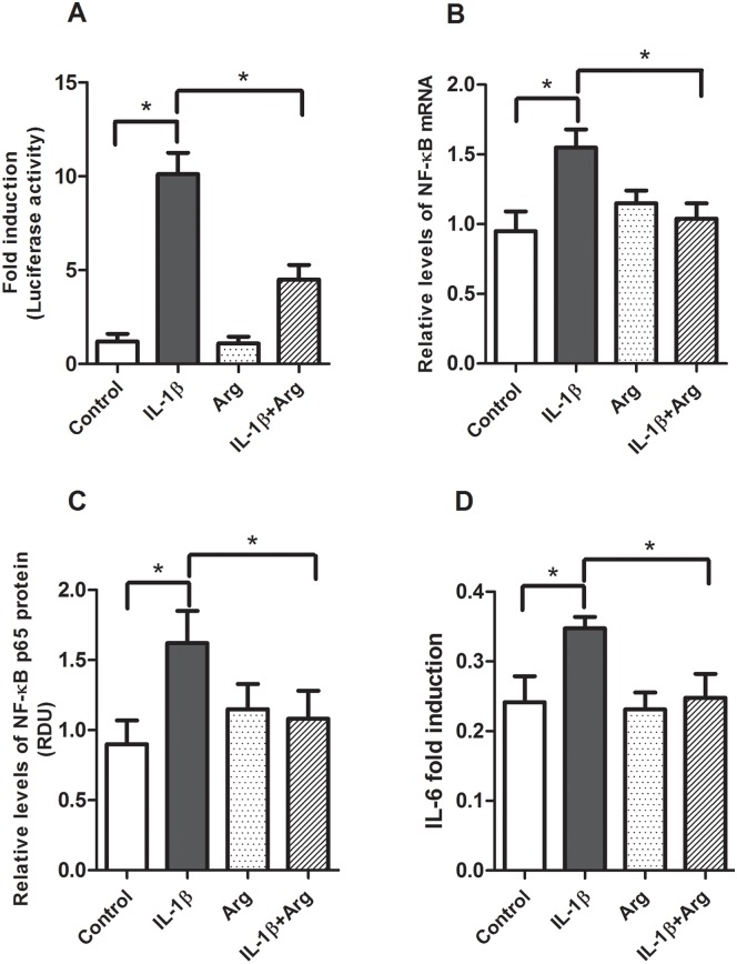
Fig 2. Effects of L-Argon IL-1β-induced NF-κB promoter activity, expression and IL-6 production.
Caco-2 cells transfected with pNF-κB-Luc vector were treated with or without IL-1β (4 ng/ml) for 4 hours after pretreatment in the absence or presence of L-Arg (5 mM) for 4 hours (Fresh medium with L-Arg was applied after removing medium for pre-treatment). Cells were harvested for luciferase activity, isolations of total RNA and protein. A: NF-κB luciferase activity data are expressed as fold induction of normalized luciferase activity. B: NF-κB mRNA levels were determined by qRT-PCR, then normalized to β-actin. C: NF-κB p65 protein levels were measured by Western blot analysis and normalized to β-actin. D: IL-6 levels in the cell culture media were measured by ELISA, normalized to total protein and expressed as fold-induction. All data are means± SE, * P<0.05. (n = 5–6 /group).

