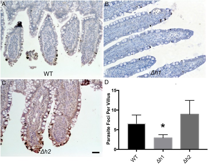
Fig 6.
Representative images of cat intestinal ileum infected with WT (A), Δh1 (B), and Δh2 (C) parasites stained with a polyclonal anti- T. gondii antibody and Streptavidin-HRP (brown). Parasites are located throughout the intestinal villi (dark deposits). Scale bar = 50μm. (D) Parasite density per villus in intestinal sections infected with WT, Δh1 and Δh2 parasites. Δh1-infected intestines showed significant reduction of parasite density (P< 0.0001, Dunn’s Multiple Comparisons test, N = 50 villi counted per section, 4 (WT, Δh2) or 6 (Δh1) independent sections counted per strain).

