Figure 3.
Physical characterization of the TPNPs.
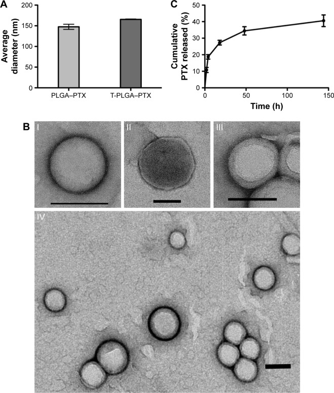
Notes: (A) Size of the PTX-loaded PLGA nanoparticles and TPNPs. (B) TEM images (n.3) of (I) a PLGA core, (II) a hCTL membrane vesicle, (III) a TPNP, and (IV) multiple TPNPs. All scale bars =100 nm. (C) Cumulative in vitro release of PTX from TPNPs (n=3).
Abbreviations: TPNPs, T-lymphocyte membrane-coated PLGA nanoparticles; PTX, paclitaxel; PLGA, poly-lactic-co-glycolic acid; PLGA–PTX, PTX-loaded PLGA nanoparticles; T-PLGA–PTX, PTX-loaded PLGA nanoparticles with hCTL membrane encapsulation; TEM, transmission electron microscopy.

