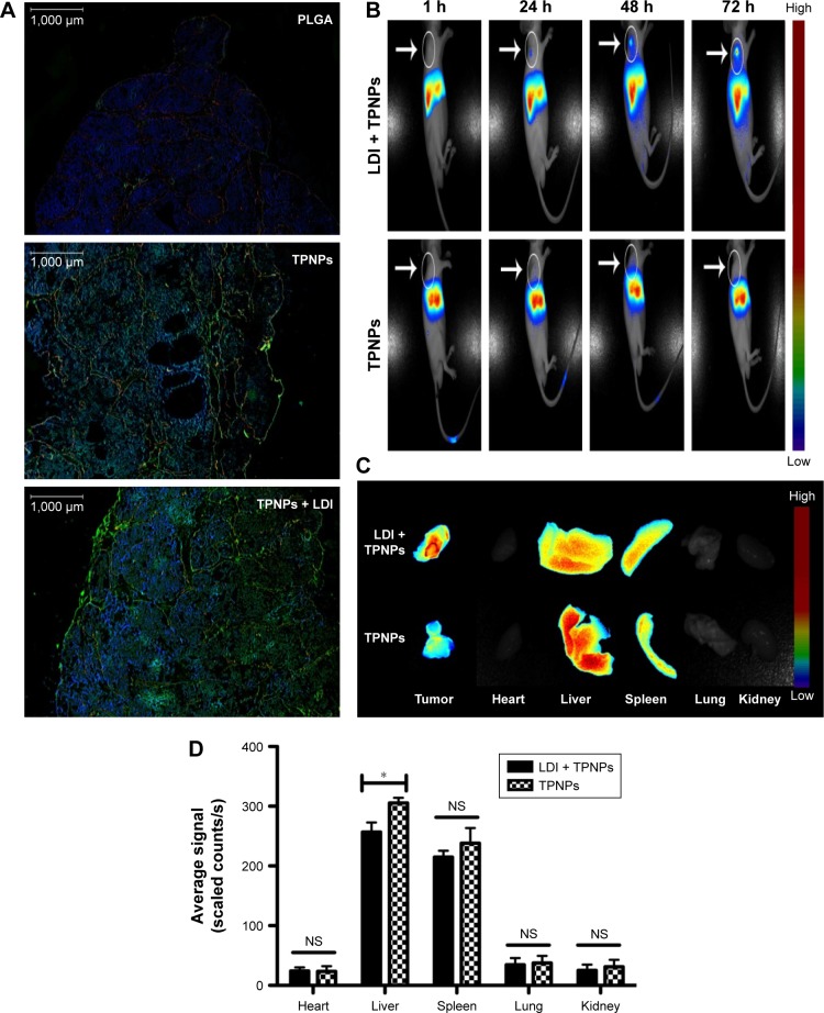
Figure 6.
In vivo targeting and biodistribution of TPNPs.
Notes: (A) Fluorescence micrographs of resected tumors. Green, DiO; red, CD31; blue, DAPI. Scale bars =1,000 μm. (B) In vivo images of real-time tumor targeting characteristics of the TPNPs, with or without LDI. (C) Ex vivo images of tumors and organs of the sacrificed mice at 96 h after the injection. (D) Biodistribution of the nanoparticles at 96 h after injection (n=3). NS, no significant difference; *P<0.05.
Abbreviations: LDI, low-dose irradiation; PLGA, poly-lactic-co-glycolic acid; TPNPs, T-lymphocyte membrane-coated PLGA nanoparticles.

