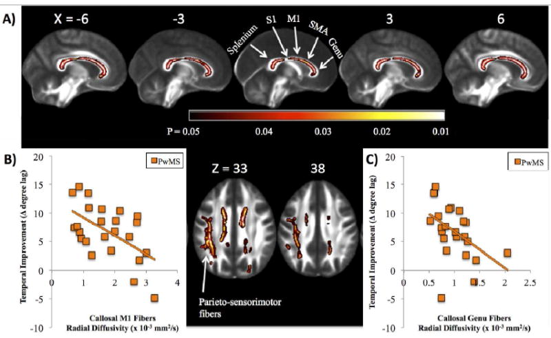
Figure 3.

A) Significant associations between temporal improvement and white matter microstructure are shown in people with MS (PwMS), with brighter colors (yellow, white) representing stronger correlation. Significant associations were localized to the corpus callosum (sagittal views: genu, body and splenium; top) and white matter connecting the posterior parietal cortices with the primary sensorimotor cortices within the left hemisphere (axial views; bottom). Results are multiple comparison-corrected and controlled for age, gender, brain volume, and EDSS. Sections of the callosum connecting specific cortical structures (splenium, primary somatosensory cortex [S1]; primary motor cortex [M1]; supplementary motor area [SMA]), and genu are localized. Callosal locations are adapted from Fling et al., 2013 [37]. Scatterplots represent individual values for PwMS displaying the significant association between temporal improvement in postural control and callosal fiber tracts connecting the B) M1: r = -0.56; P = 0.004, as well as the C) genu: r = -0.47; P = 0.01.
