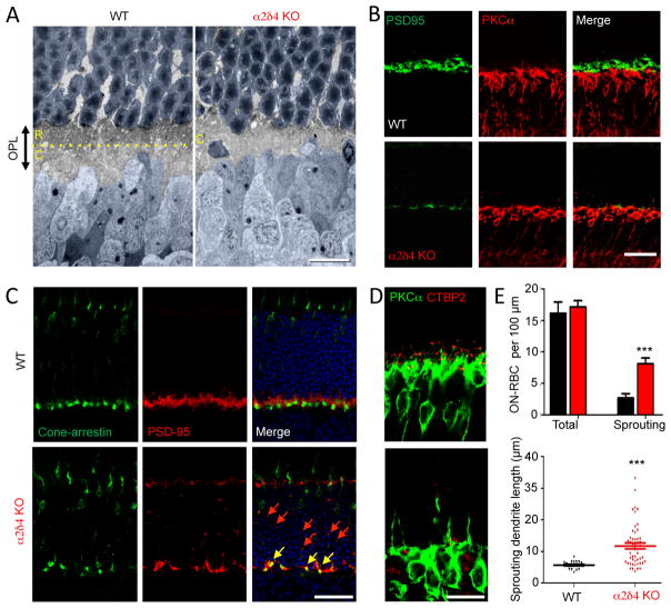
Figure 6. α2δ4 ablation causes deficits in elaboration of rod axonal terminals.
A, Ultrastructural analysis of the outer plexiform layer (OPL) organization. Rod photoreceptor axons terminate in the outer aspect of the OPL (R), whereas cones form pedicles in the inner sublamina (C), separated by the yellow dotted line. Nuclei of photoreceptors and ON-BC are painted in blue. B, Loss of rod synaptic terminals identified by PSD95 staining in the OPL region of α2δ4 knockout retinas. C, Preservation of cone axonal terminals in the OPL region of α2δ4 knockout retinas and axonal retraction of rod terminals to the outer nuclear layer (ONL). Cone pedicles were identified by overlap between cone arrestin and PSD95 staining (yellow arrows). Remaining rod terminals are seen as small PSD95 positive (cone arrestin negative) puncta scattered in the OPL and ONL (red arrows). D, Sprouting ON-RBC dendrites in α2δ4 KO retinas. E, Quantification of dendritic overextension (sprouting) of ON-RBCs in α2δ4 KO comparison relative to WT. Six to eight regions of retina with different eccentricities from 3 independent mice for each genotype were used. Error bars are SEM values, ***p<0.001, t-test. All scale bars in A–E are 25 μm.

