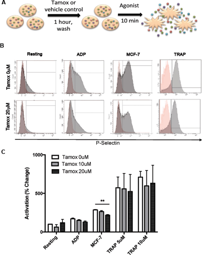
Figure 2. Tamoxifen inhibits platelet activation.

To measure platelet activation, platelets were isolated from healthy donors, pretreated with 10–20 µM of tamoxifen or vehicle control, washed, and exposed to agonists (ADP, MCF-7 tumor cells or TRAP). (A). P-selectin surface expression, a marker of activation, was determined by flow cytometry. Representative histograms are shown, with P-selectin stained platelets (gray) overlaid onto platelets stained with isotype control antibodies (red) (B). Quantified results are shown in C. Bars indicate SEM. P<*0.01 compared to resting control unless otherwise indicated by ANOVA, n=3–6 independent replicates per treatment group.
