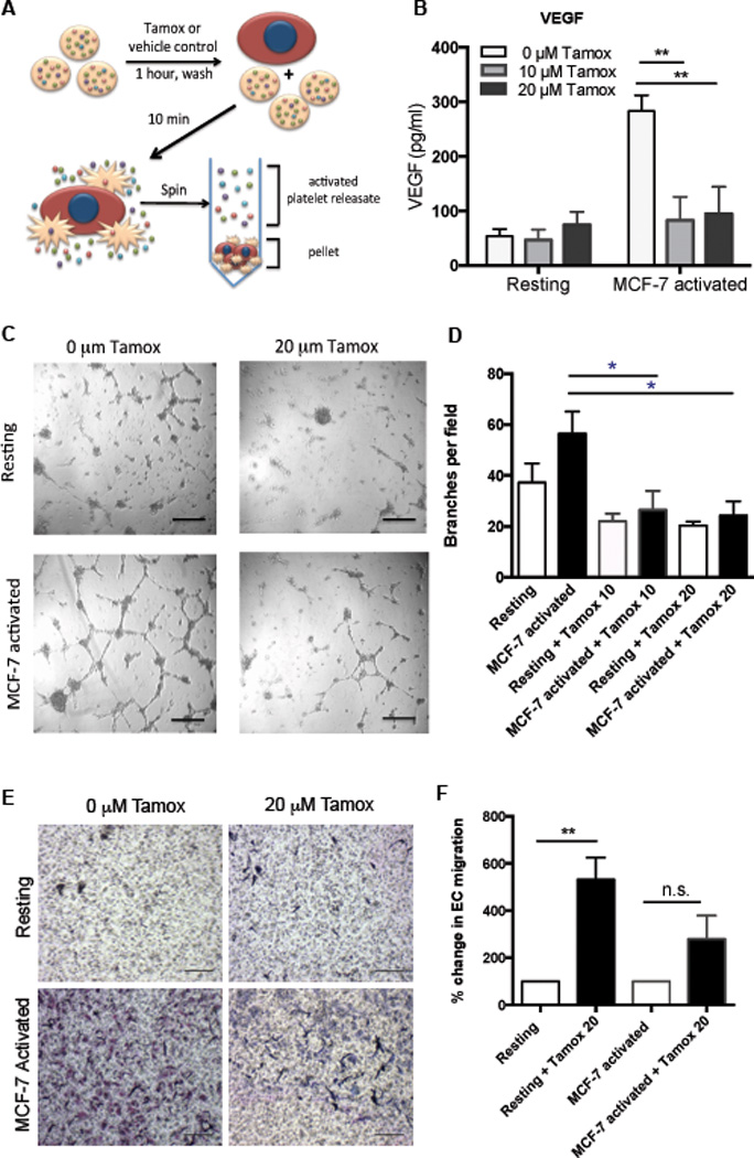
Figure 3. Tamoxifen decreases the angiogenic potential of platelets.

Platelets were pretreated with 0, 10, or 20 µM tamoxifen, washed, and activated with MCF-7 tumor cells or left unactivated (resting) to generate releasates (A). VEGF was quantified in releasates by ELISA (B). Capillary tube formation in HUVECs was assessed following 6 hours of exposure to platelet releasates and quantified as the average number of branch points per field of view (D), with representative images shown (C). Endothelial migration in the presence of resting or MCF-7-activated releasates generated from tamoxifen (20 µM) or control treated platelets was quantified (D-E). Representative images are shown (E) and data from all replicates are quantified as the average number of migrated HUVECs per field (D). Bars indicate SEM. P<*0.05, **0.01 by ANOVA, n=3 independent replicates per treatment group. Scale bars represent 100 µm.
