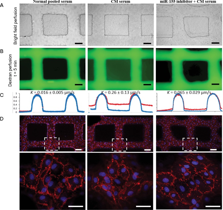
Figure 7.
Human umbilical vein endothelial cell (HUVEC) microvessels perfused with sera from children with cerebral malaria (CM) illustrate greater dextran leak versus sera control, which can be prevented with preincubation with miR-155 antagomir. (A) Bright field image of human sera perfused through the microvessels: normal pooled sera (left panel), CM sera (middle panel) and microvessels pretreated with antagomir-155 prior to CM sera (right panel) (scale bar: 100 μm). (B). Fluorescence image of microvessels perfused with 40 kDa dextran (as a marker of microvascular leak) after 5 min of perfusion, after different sera treatments. (C) Line cut of intensity profile of dextran-perfused microvessels in B at t = 1 min (blue line) and t = 5 min (red line). The permeability K of three conditions was determined to be 0.016 ± 0.005 μm/s (normal sera), 0.26 ± 0.13 μm/s (CM sera) and 0.065 ± 0.029 μm/s (antagomir-155 + CM sera). (D) Z-stack projection of confocal fluorescence images of microvessels after serum treatment and dextran perfusion, stained with CD31 (cell junction) and Hoechst 33342 (nuclei) (scale bar: 100 μm). Bottom panels: zoomed-in view of dashed square box (scale bar: 50 μm). Confocal images illustrate no differences in PECAM1/CD31 cell junctions despite significant differences in dextran leak. The assay was repeated with a different CM sample and yielded similar results (n = 2 biological replicates).

