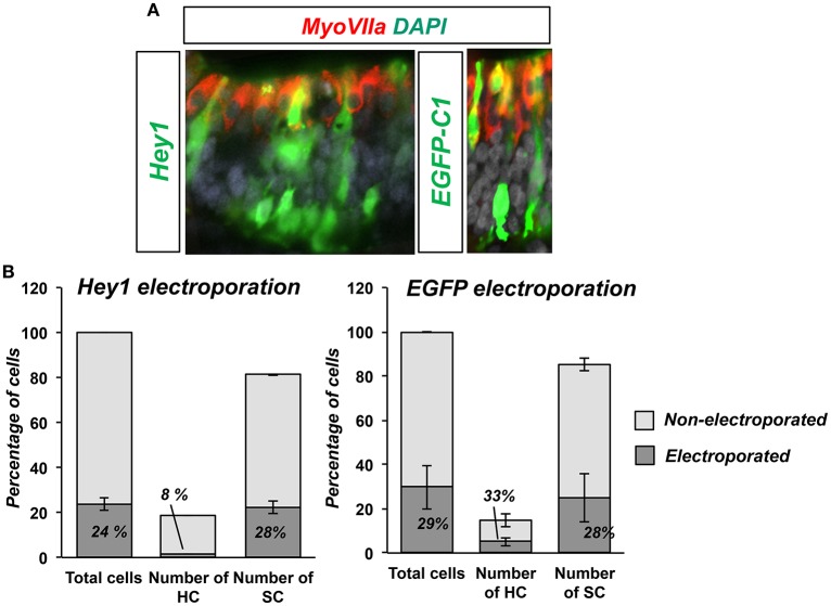
Figure 5.
Hey1 prevents HC formation in ovo. (A) E3.5 chicken embryos were electroporated with Hey1 (left image) or EGFP-C1 (right image) and then sectioned after 3 days of incubation (E3.5+3). Electroporated cells in the macula sacularis were found mainly in the SC layer, and very few developed as HCs. Control electroporation with EGFP-C1 (E3.5+3) is shown on the right. (B) Hey1 electroporation biased electroporated cells toward supporting cell fate. The fraction of HCs that were electroporated (8%) was smaller than that of SCs (28%), similar to the efficiency of the electroporation (24%). Bars represent the number of cells counted in two consecutive frames of electroporated macula sacularis, from three independent embryos (n = 3). Electroporation with EGFP-C1 did not show any bias for either HCs or SC.

