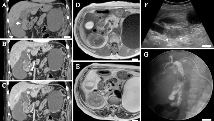
Figure 1.
Radiographic images of the extrahepatic bile duct tumor. A-C: Coronal plane of computed tomography (CT) images. A precontrast CT image (A) shows the extrahepatic bile duct mass (arrow) and the dilated gallbladder (arrowhead). The intrahepatic and extrahepatic bile ducts are dilated. A large renal cyst is present in the left kidney. An arterial phase CT image (B) shows that the mass has slight and heterogeneous enhancement. A delayed phase CT image (C) shows that the mass is further enhanced. D-E: Magnetic resonance imaging shows that the mass is heterogeneous and somewhat hypointense on a T1-weighted image (D) and partially hyperintense (arrow) on a T2-weighted image (E). The hyperintense area probably corresponds to the largest tumor. F: Sagittal plane of the ultrasonography image. An intraductal nodule 7 cm in length with dilatation of the extrahepatic bile duct is seen. The nodule includes a hypoechoic lesion (arrowheads) that probably corresponds to the largest tumor of four polypoid lesions. G: Percutaneous transhepatic cholangiography. A filling defect is seen in the extrahepatic bile duct. The paucity of the transverse portion of the duodenum suggests intestinal malrotation. White bar, 2 cm in A, D-G.

