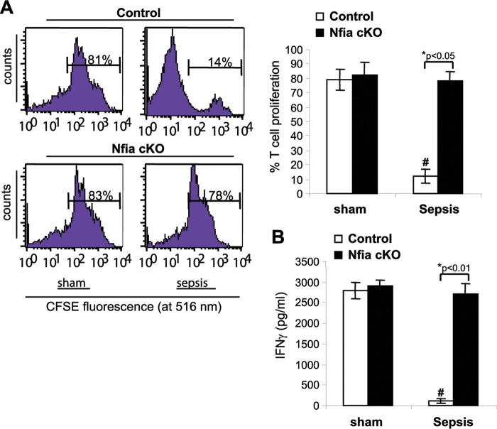
FIG 7.
Gr1+ CD11b+ cells from late-sepsis NFI-A conditional knockout mice do not suppress T cell activation or proliferation. A coculture of Gr1+ CD11b+ cells and T cells was used to assess the immunosuppressive function of Gr1+ CD11b+ cells. Spleen CD4+ T cells were isolated from naive wild-type mice and labeled with the fluorescent dye CFSE for 10 min at room temperature. Bone marrow Gr1+ CD11b+ cells isolated from sham treatment or late-sepsis mice were then cocultured (at 1:1 ratio) with CD4+ T cells, and the culture was stimulated with anti-CD3 plus anti-CD28 antibodies (1 μg/ml each). (A) After 3 days, CD4+ T cell proliferation was determined by the stepwise dilution of CFSE dye in dividing CD4+ cells by flow cytometry. Histograms of gated T cells (left) and quantitative analysis of cell proliferation (right) are shown. Percentages of cell proliferation were calculated as follows: percent cell proliferation = 100 × (count from CD4+ T cell + Gr1+ CD11b+ cell culture/count from CD4+ T cell culture alone). (B) The culture supernatants were analyzed for IFN-γ production by ELISA. Data are expressed as means ± SD of results from 5 mice per group pooled from three experiments. *, P value (control versus cKO, Student's t test); #, P < 0.05 (sham treatment group versus sepsis group, ANOVA). cKO, conditional knockout.

