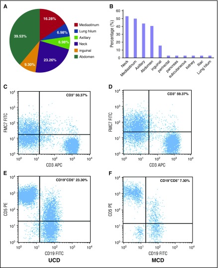
Figure 1.
Lymph node involvement by location and immunophenotypic expression (CD3+ and CD5+/CD19+) in patients with UCD and iMCD subtypes of CD. (A) The distribution of lymphadenopathy among patients with HIV-negative UCD. (B) The locations of coexistent lymphadenopathies among patients with iMCD; (C,E) Flow cytometry images of CD3+ and CD5+/CD19+ in UCD; (D,F) flow cytometry images of CD3+ and CD5+/CD19+ in iMCD. FITC, fluorescein isothiocyanate; PE, phycoerythrin.

