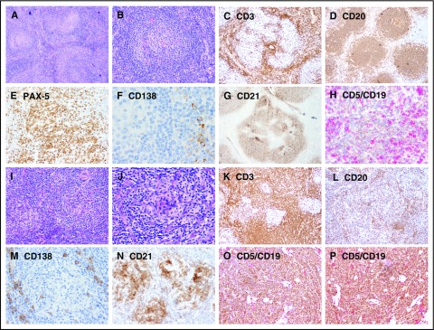
Figure 2.
Representative images showing immunohistochemical expression in patients with UCD and iMCD. (A-H) UCD case with B-cell–rich germinal centers and increased CD5+/CD19+ B cells, whereas CD3+ small T cells are relatively sparse. (I-P) iMCD case with dense T cells in the interfollicular regions and decreased CD20/PAX-5 B cells and CD5+/CD19+ B cells. Few polyclonal CD138+ PCs are present around the nodules in both UCD and iMCD. Original magnification ×100 for panels A-D,I-L and ×200 for panels E-H,M-P.

