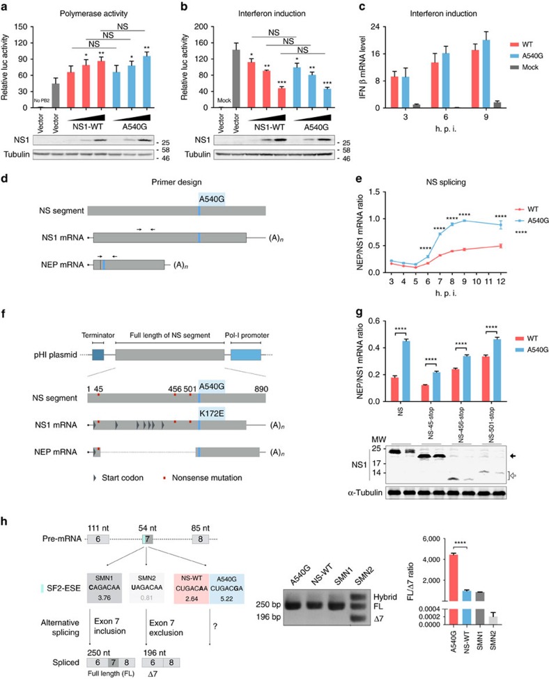
Figure 2. A540G enhances NS splicing ratio.
(a) HEK293T cells were transfected with RNP complexes (PB2, PB1, PA and NP) and vector, different amounts of H7N9-NS1-WT or H7N9-NS1-A540G (K172E) mutant NS1 plasmids, together with firefly (pYH-Luci for RNP activity) and Renilla (control) luciferase reporters, for 16 h. (b) HEK293T cells were transfected with vector, different amounts of H7N9-NS1-WT or H7N9-NS1-A540G (K172E) NS1, together with IFN-β and Renilla reporters. At 8 h post transfection, cells were infected or mock-infected with SeV and luciferase activities were analysed after cultured overnight. (c) IFN-β mRNA induction in A549 cells mock infected or infected with rH9N2-WT or rH9N2-NS-A540G viruses (multiplicity of infection (MOI)=1) was analysed by RT–qPCR. (d) The black arrowheads indicate primers for detection of NS1 and NEP mRNA used in e. (e) RNA from A549 cells infected with rH9N2-WT or rH9N2-NS-A540G viruses (MOI=1) was analysed by RT–qPCR. The splicing ratio was calculated by the ΔCt method. (f) Potential in-frame start codons in the H7N9 NS1 gene are shown as grey triangles. The three plasmids used in g include different stop codons (red squares), NS-45-stop aims to truncate the NS1 protein in N-terminal and for NS-456-stop and NS-501-stop, to abort translation before amino acid 172 (nucleotide 540). (g) HEK293T cells were transfected with pHI-H7N9-NS or its mutants, as indicated, together with H7N9 RNP complexes. The splicing ratio of NS mRNAs was analysed by RT–qPCR. Expression of WT and mutant NS1 proteins was analysed by immunoblotting. The solid arrowhead indicates proteins translated from the second in-frame start codon, while the unfilled arrowhead indicates truncated NS1 proteins ending before residue 172. (h) Schematic illustration of SMN mini-gene splicing assay. The verified ESE motif in exon-7 is shown in blue. SMN1 is a wild-type ESE, whereas SMN2 is dysfunctional. The SF2-ESE motif score (ESEfinder) is shown under the sequence. If the ESE is functional, exon 7 would be included during alternative splicing (full length, FL). Otherwise, it would be excluded (−7), leading to a shorter spliced product. RNAs from HEK293T cells transfected with pSMN plasmids containing the indicated ESE motifs were analysed. The ratio of FL to −7 mRNA from the SMN gene was estimated by RT–qPCR. Error bars represent mean±s.d. (n=3). Statistical significance was analysed by (e,c) two-way analysis of variance with Bonferroni post test or by (a,b,g,h) Student's t-test: *P<0.05, **P<0.01, ***P<0.001 and ****P<0.0001; NS, not significant. Symbols above graph bars or plotted points indicate the statistical significance of the comparison between WT and A540G groups.

