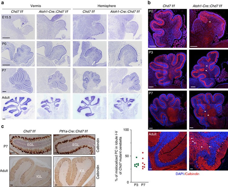
Figure 2. Genetic ablation of Chd7 in GNPs leads to cerebellar hypoplasia and Purkinje cell ectopia.
(a) Nissl staining of E15.5, P0, P7 and adult cerebella including vermis and hemisphere from [Chd7f/f] and [Atoh1-Cre::Chd7f/f] mice. Left side in each panel is the anterior part of cerebella. Scale bars, 100 μm. (b) Co-immunostaining of Calbindin (red), a marker for Purkinje cells (PCs) in P0, P3, P7 and adult WT [Chd7f/f] and Chd7 mutant [Atoh1-Cre::Chd7f/f] cerebella. Cellular nuclei are counterstained with DAPI (blue). Arrowheads indicate clusters of mislocalized PCs. Scale bars, 200 μm. Quantification of the percentage of mislocalized PCs among all PCs in anterior lobule I-V is shown in the left panel. Cerebellar sections from three independent P3 and six P7 Chd7 mutant mice were counted. (c) Immunostaining of Calbindin in cerebella of P7 and adult WT [Chd7f/f] and Chd7 mutant [Ptf1a-Cre::Chd7f/f] mice. Sections were counterstained with haematoxylin. Scale bars, 100 μm.

