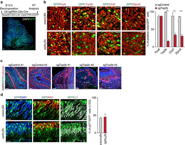
Figure 7. Genetic ablation of Top2b leads to phenotypic changes analogous to Chd7-deficient cerebella.
(a) Immunostaining of GFP (green) on P7 cerebellum from Rosa26-CAG-LSL-Cas9-P2A-EGFP mouse that was electroporated at E13.5 with a plasmid carrying sgRNA and Cre. Section is counterstained with DAPI (blue). Scale bar, 100 μm. The experimental scheme is shown in the top panel. (b) Immunostaining of GFP and Pax6, Top2b, Chd7 and Zfpm2 on P7 cerebella from Rosa26-CAG-LSL-Cas9-P2A-EGFP mouse that was electroporated at E13.5 with a plasmid construct expressing Cre and sgRNA against Top2b (sgTop2b) and a control sequence (sgControl). Quantification of positive cells among GFP+ cells is shown in the right panel. Two and three pups were analysed for sgControl and sgTop2b, respectively. P=8.567E-06 (for Top2b), P=0.0037 (for Chd7), P=0.00046 (for Zfpm2). Scale bars, 20 μm. (c) Immunostaining of GFP (green) and Calbindin (red) of P7 sgControl- and sgTop2b-electroporated cerebella. Arrowheads highlight mislocalized Purkinje cells. Scale bars, 100 μm. (d) Immunostaining of GFP (green), Ki67 (red) and p27 (white) of the EGL from P7 sgControl- and sgTop2b-electroporated animals. Quantification of p27+ cells among GFP+ cells was shown in the right panel. Scale bars, 20 μm.

