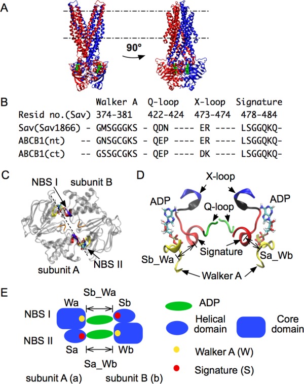
Figure 1.

Architecture of ABC exporters. The X-ray structure of Sav1866 (PDB ID: 2HYD) is shown with bound ADP molecules. A: Side views of the whole structure. The two subunits are respectively rendered in blue and red. Membrane boundaries are indicated by dashed lines: upper, extracellular side; lower, intracellular side. ADP is shown in green. B: Sequence alignment showing the conservation of motifs in Sav1866 (Sav) and P-glycoprotein (Pgp, ABCB1). nt, ct: N-terminal and C-terminal side in the heterodimer Pgp (Sav is a homodimer). C: Top view of NBD with motifs highlighted. Two NBDs (subunits A and B) form two nucleotide binding sites, NBS I and NBS II. D: Side view of NBD motifs. The X-loop and the signature motif are connected by a short loop, colored in black. E: Scheme of the two NBSs, highlighting the two binding distances, Sb_Wa and Sa_Wb. Sa, Sb: signature motif from subunit A(a) and B(b). Wa, Wb: Walker A from the two subunits. Sb_Wa: distance between Sb and Wa, at NBS I. Sa_Wb: distance between Sa and Wb, at NBS II.
