
Figure 2.
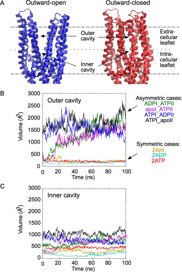
Two different conformations of the TMDs. A: Outward-open and outward-closed conformation of TMDs, taken at t = 100 ns in the simulations of the ATPI_ADPII and 2ATP states, respectively. Membrane boundaries are indicated by dashed lines. B, C: Volume of the cavity formed by the TMDs at the level of the extracellular leaflet (B) and intracellular leaflet (C) for different nucleotide occupancy states.