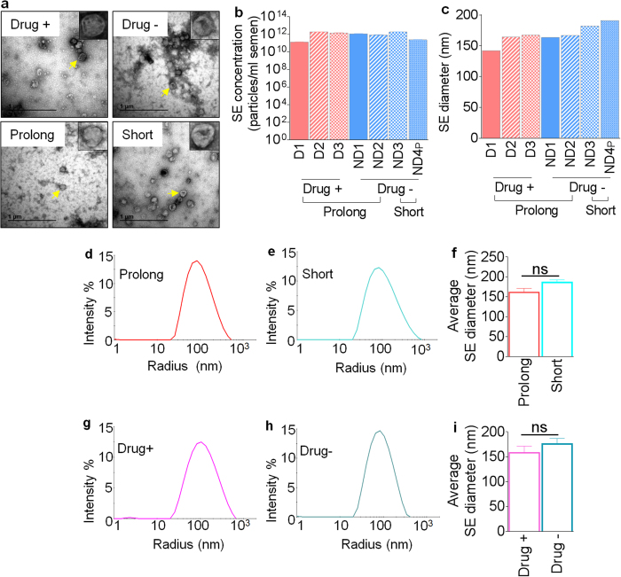
Figure 1. Physical properties of SE isolated from diverse conditions.
(a) Representative TEM images showing SE morphology from donors who used illicit drugs (top left) or did not use illicit drugs (top right) and following prolonged (bottom left) or short (bottom right) term freezing of semen. The insets on each image show the zoomed image of a single vesicle. Yellow arrowheads indicate the vesicle in each field used for the zoomed image. Scale bars are 1 μm. (b) Exosome concentrations were quantified by NTA and averaged from three measurements. Concentrations were calculated per ml of semen. (c) Comparison of SE size in diameter (nm) by length of storage or donor illicit drug use. (d,e) Representative histograms showing SE distribution intensity by radius (nm) following prolonged (n = 5) or short-term freezing (n = 13) of semen. (f) Average SE size in diameter (nm) by length of storage. (g,h) Representative histograms showing distribution intensity of SE from donors who used (n = 3) illicit drugs or donors who did not use illicit drugs (n = 15). (i) Average SE size in diameter (nm) by donor illicit drug use. For statistical analysis, samples were grouped into “length of the freezing” and “drug use” and analyzed comparing short to prolong or drug− to drug+ respectively for F and I. Significance was determined by student’s t test. Differences with p values of 0.05 are considered significant. Error bars are standard errors of the mean. ns = not significant.

