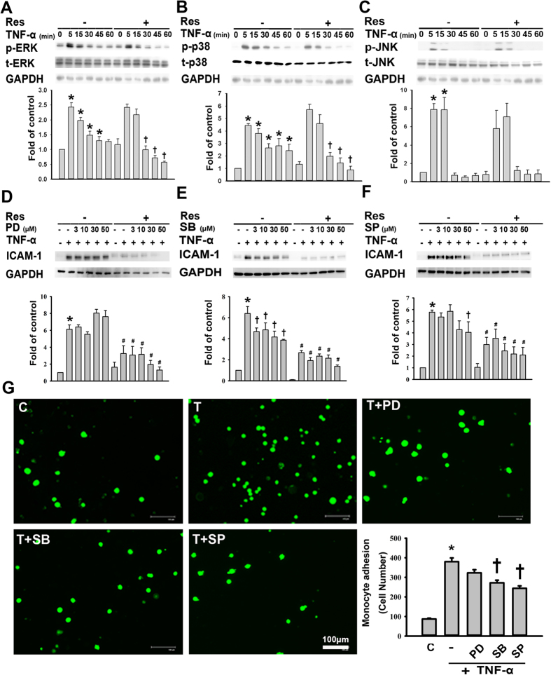
Figure 2. The resveratrol-mediated reduction in TNF-α-induced ICAM-1 expression is partly dependent on the inhibition of p38 phosphorylation.
(A–C) The effects of resveratrol treatment on the phosphorylation of ERK1/2, p38, or JNK in TNF-α-treated HUVECs. HUVECs were incubated for 24 h with or without 50 μM resveratrol; then, the cells were incubated with 3 ng/mL of TNF-α for the indicated time, and aliquots of cell lysates containing equal amounts of protein subjected to immunoblotting with antibodies against (A) p-ERK1/2 and t-ERK1/2, (B) p-p38 and t-p38, (C) or p-JNK and t-JNK. (D–F) HUVECs were incubated for 23 h with or without 50 μM resveratrol, with or without the subsequent addition of the indicated concentrations of (D) PD98059 (an ERK1/2 inhibitor), (E) SB203580 (a p38 inhibitor), or (F) SP600125 (a JNK inhibitor) for 1 h in the continued presence of resveratrol. This was followed by incubation with or without TNF-α for 4 h, and then the cell lysates were analyzed for ICAM-1 expression by Western blot. The data are expressed as a fold of the control value and are shown as the mean ± SEM of three separate experiments. GAPDH was used as the loading control. Blots are cropped for clarity; full blots are shown in the Supplementary file. (G) Representative fluorescence images showing the effects of MAPK inhibitors on the TNF-α-induced adhesion of fluorescein-labeled U937 cells to HUVECs. Cells were left untreated or were pretreated for 1 h with PD98059 (30 μM), SB203580 (30 μM), or SP600125 (30 μM). Then, they were treated with 3 ng/mL TNF-α for 4 h in the continued presence of the inhibitor. BCECF-AM-labeled U937 cells were added to HUVECs and incubated at 37 °C for 45 min. The adherent cells were imaged by fluorescence microscopy. Bar = 100 μm. The number of U937 cells bound per high power field in six randomly selected images was counted. The data are expressed as the mean ± SEM of three separate experiments. *P < 0.05 compared with the untreated cells. †P < 0.05 compared with the TNF-α-treated cells.

