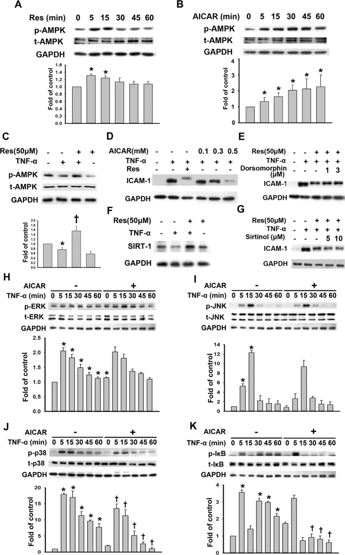
Figure 4. Resveratrol-mediated decreases in TNF-α-induced ICAM-1 expression and partly dependent on AMPK phosphorylation.
(A) Representative immunoblot analysis of the time-dependence of the resveratrol-mediated phosphorylation of AMPK (p-AMPK) in HUVECs. The density of the p-AMPK bands was quantified and normalized to the protein loading control GAPDH, and the mean ± SEM are shown (n = 3). (B) Immunoblot analysis of the time-dependence of AICAR (an AMPK activator)-mediated AMPK phosphorylation. (C) Western blot analysis of the p-AMPK expression. HUVECs were preincubated for 24 h with 50 μM resveratrol and were then treated with 3 ng/mL TNF-α for 4 h. (D) Western blot analysis of ICAM-1 expression. HUVECs were incubated with the indicated concentrations of AICAR for 24 h and then with 3 ng/mL TNF-α for 4 h in the continued presence of AICAR; then, the ICAM-1 protein expression in cell lysates was measured by Western blot. (E) Western blot analysis of ICAM-1 expression. HUVECs were incubated with the indicated concentrations of Dorsomorphin (an AMPK inhibitor) and 50 μM resveratrol for 24 h and then with 3 ng/mL TNF-α for 4 h; then, the ICAM-1 protein expression in the cell lysates was measured by Western blot. (F) Western blot analysis of SIRT-1 expression. HUVECs were preincubated for 24 h with 50 μM resveratrol and were then treated with 3 ng/mL TNF-α for 4 h. (G) Western blot analysis of ICAM-1 expression. HUVECs were incubated with the indicated concentrations of Sirtinol (a SIRT-1 inhibitor) and 50 μM resveratrol for 24 h and then with 3 ng/mL TNF-α for 4 h; then, the ICAM-1 protein expression in the cell lysates was measured by Western blot. (H-K) The effects of AICAR treatment on p-ERK1/2, p-JNK, p-p38, and p-IκB in TNF-α-treated HUVECs. HUVECs were incubated for 24 h with or without 0.5 mM AICAR, and then the cells were incubated with 3 ng/mL of TNF-α for the indicated time. Cell lysates were subjected to immunoblotting with the antibodies against (H) p-ERK1/2 and t-ERK1/2, (I) p-p38 and t-p38, (J) p-JNK and t-JNK, or (K) p-IκB and t-IκB. *P < 0.05 compared with the untreated cells. †P < 0.05 compared with the TNF-α-treated cells at the same time point. Blots cropped for clarity; full blots are shown in the Supplementary file.

