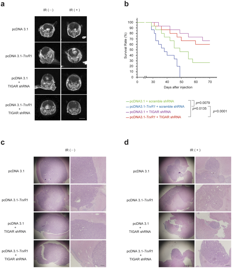
Figure 6. TIGAR abrogation increases the radiosensitivity of TrxR1-overexpressing glioma in vivo.
(a) Axial T1-weighted images of tumour-bearing mice. At the end of brain-focalized radiotherapy, contrast enhanced MRI images of the animal brains were obtained. Left panels: sham IR, Right panels: 20-Gy fractionated IR (scale bar = 6 mm). (b) The Kaplan-Meier survival analysis of nude mice inoculated with U-87MG cells as indicated. Each group was composed of 15 female BALB/c nude mice. (c,d) H&E staining of xenografts with or without irradiation was performed post 20-Gy fractionated radiotherapy. Left panels: 40x magnification (scale bar = 500 μm), Right panels: 100x magnification (scale bar = 100 μm).

