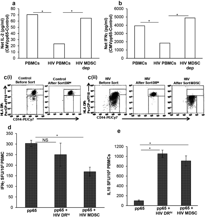
Figure 2. In vitro expanded HIV MDSC are functionally similar to MDSC in HIV-infected individuals and modulate CMV specific T cell response.
PBMCs of CMV(+) HIV(−) donors were cultured without (PBMCs) or with inactivated HIVBaL (HIV PBMCs) (p24 Ag, 12 ng/107 cells). After 5 days, cells were stained with anti-CD11b, -CD33, CD14, HLA DR. (a and b) CD14+HLA DR−/lo MDSC were depleted from HIV PBMCs. HIV PBMCs, HIV PBMCs and MDSC depleted HIV PBMCs (HIV MDSC dep) were cultured with or without CMVpp65 for 24 and 48 hrs. The amounts of IL-2 (a) and IFNγ (b) were determined in culture supernatants at 24 and 48 hrs, respectively. Net IL-2 or IFNγ was calculated as CMVpp65 – Control. (c and d) PBMCs were cultured and stained as above, DRhi and MDSC were sorted. (ci and cii) Representative dot plots show HLA DR vs CD14 in control cells before sort and after sort (Left panel) and in HIVBaL treated cells before sort and after sort (Right panel). Cells isolated are >98% pure. (d and e) Isolated HIV DRhi and MDSC (5 × 10⁴) were cultured overnight with autologous freshly isolated PBMCs (1 × 10⁵) on ELISPOT plates with or without CMVpp65 to determine frequency of (d) IFNγ producing cells and (e) IL-10 producing cells. Histograms are presented as mean+/−SD; n = 3 donors; *p < 0.05.

