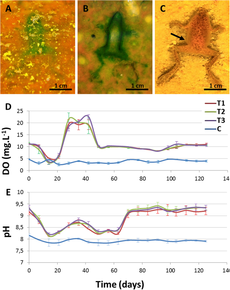
Figure 1. Taphonomic alterations of frog carcasses over a mat.
(A) and (B), outlines of removed frog bodies, which are greened by local stimulation of the phototrophic mat populations. In (B), the thickness of the frog contour is due to incipient sarcophagus formation. Experiments in (A) and (B) were conducted at day 3 and day 7, respectively. (C) Frog in the control showing a darkened outline of the sediment that is in direct contact with the body (day 7). (D) and (E), variations of DO and pH in water from the tanks with mats (T1-3) and the control (C) over the course of the experiment.

