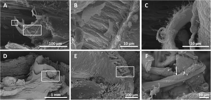
Figure 5.
SEM photos of muscle (A–C) and connective tissue (D–F) in the frog in the mat at day 1080. (A) Femoral muscle that was ripped during the preparation shows different layers that have been magnified in (B) and (C). (D) Femoral knee articulation of a frog inset in the box. (E) Fibrous fibres of tendons from the same area, magnified in (F). The arrow highlights the striping of collagenous fibres.

