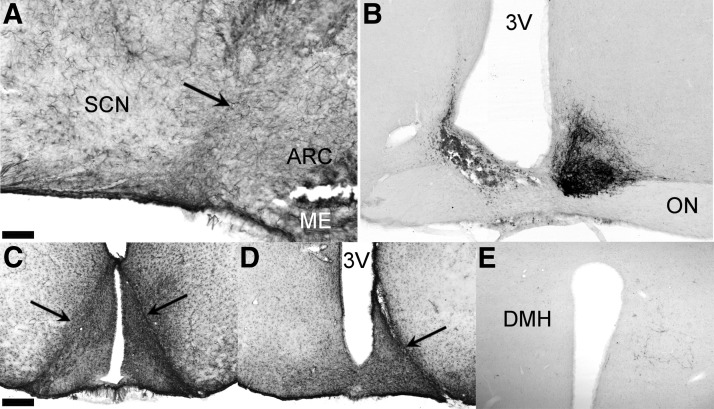
Figure 1.
Representative sagittal and coronal sections of the hypothalamus illustrating the 45° angle knife cut and its effect established through GFAP and VIP staining. A, Sagittal GFAP-stained section just lateral to the third ventricle with minimal glial damage around the site of incision indicated by a black arrow. B, Unilateral SCN lesion in combination with a contralateral knife cut with VIP staining showing the contralateral SCN intact. C, Shows GFAP staining of the most caudal reach of the knife isolating the ARC from the SCN. D, Unilateral RC-cut isolating the ARC contralateral to the SCN lesion shown in B. E, Unilateral VIP innervation of the DMH on the side of the RC-cut demonstrating effective unilateral innervation as compared with the loss of innervation on the SCN-lesioned side (left). Scale bar, 100 μm (A), 90 μm (B), 250 μm (C and D), and 130 μm (E). ME, median eminence; 3V, third ventricle.

