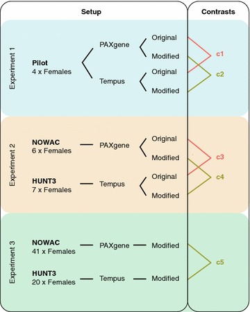
Fig. 1.

Study design. Differences in gene expression between PAXgene and Tempus were investigated in three experiments. In experiment 1 (light blue), four volunteers donated blood samples on PAXgene and Tempus tubes and RNA was isolated with both the original protocols and the modified protocol. Paired statistical analyses identified differences between PAXgene and Tempus for the original protocols (contrast 1) and for the modified protocol (contrast 2). In experiment 2 (light orange), RNA was isolated with both the original protocols and the modified protocol from two different biobanks—NOWAC and HUNT3—which had samples on PAXgene and Tempus tubes, respectively. Non-paired statistical analyses identified differences between PAXgene and Tempus for the original protocols (contrast 3) and for the modified protocol (contrast 4). In experiment 3 (light green), RNA was isolated with the modified protocol from a larger set of samples from NOWAC and HUNT3 and a non-paired analysis was performed (contrast 5). Comparisons between PAXgene and Tempus based on the original protocols are highlighted in orange (contrasts 1 and 3), whereas comparisons based on the modified protocol are highlighted in olive green (contrasts 2, 4, and 5)
