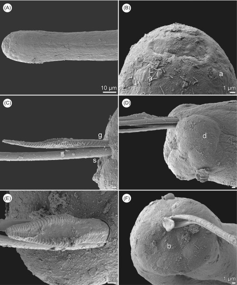
Figure 3.
Philometra rara n. sp., scanning electron micrographs. (A) Anterior end of male; (B) cephalic end of male, dorsoventral view; (C) gubernaculum, lateral view; (D) caudal end of male, lateral view; (E) gubernaculum, dorsal view; (F) caudal end of male, apical view. Abbreviations: (a) submedian pair of outer cephalic papillae; (b) dorsal caudal papilla; (c) group of four adanal papillae; (d) caudal mound; (g) gubernaculum; (s) spicule.

