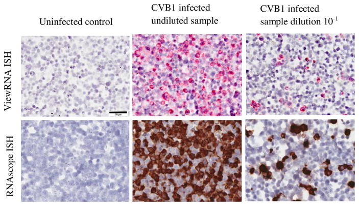
Fig. 4.
Detection of CVB1 in infected A549 and in uninfected A549 cells (FFPE) using two different commercially available ISH (ViewRNA and RNAscope) methods. Example micrographs of uninfected control, CVB1 infected undiluted sample and CVB1 infected dilution 10−1 are shown. 40× magnification. Scale bar = 50 μm.

